
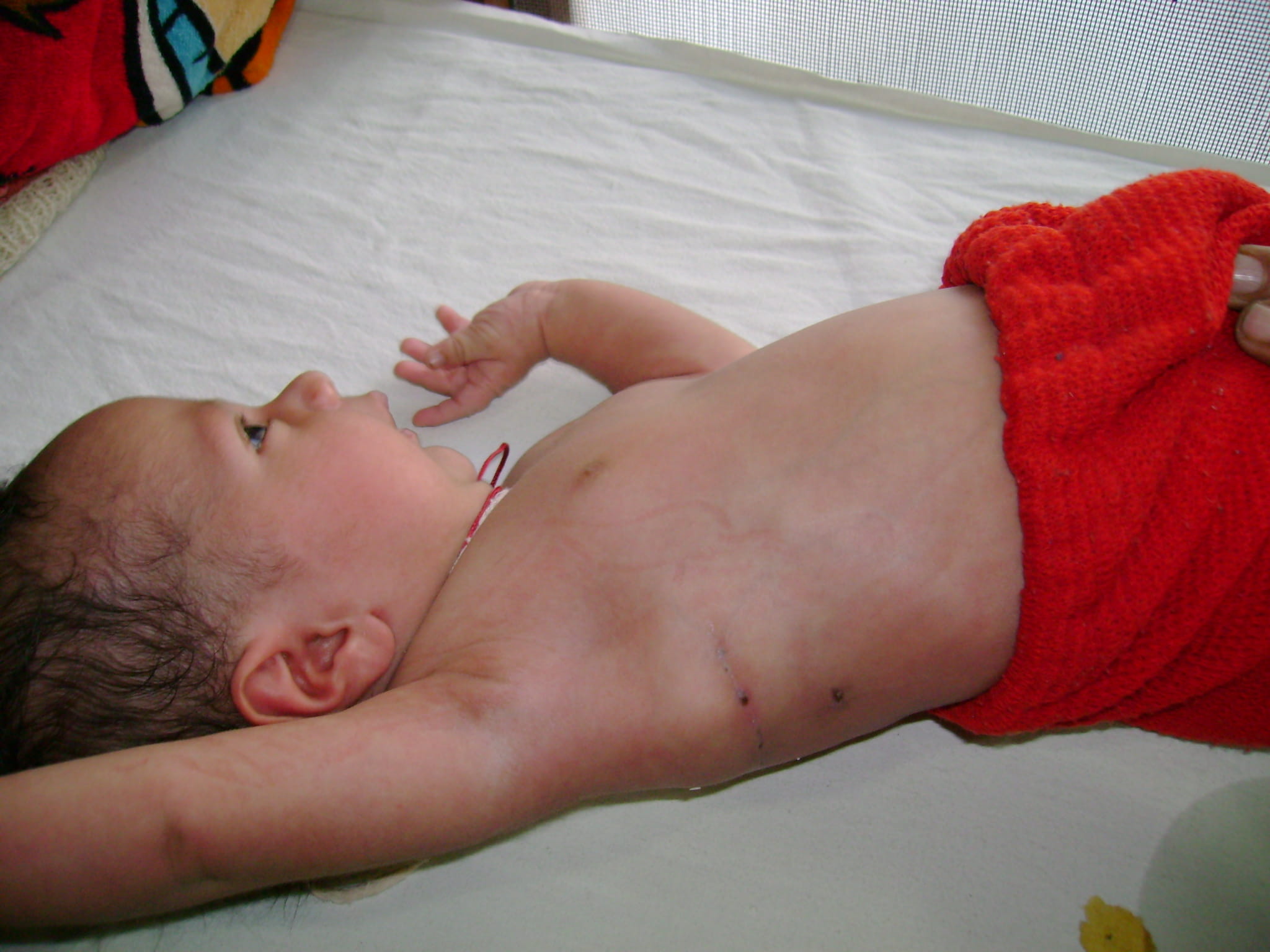
Tracheoesophageal Fistula and Oesophageal Atresia
Tracheoesophageal fistula is an abnormal connection in one or more places between the oesophagus (the tube that leads from the throat to the stomach) and the trachea (the tube that leads from the throat to the windpipe and lungs). Normally, the oesophagus and the trachea are two separate tubes that are not connected. Tracheoesophageal fistula is also known as TO fistula or simply TOF. TO fistula is a birth defect, which occurs in 1 in 5,000 births, and occurs as a foetus is forming in its mother's uterus. When a baby with a TO fistula swallows, the liquid can pass through the abnormal connection between the oesophagus and the trachea. When this happens, liquid gets into the baby's lungs, which can cause pneumonia and other problems.
What is oesophageal atresia?
TO fistula often occurs with another birth defect known as oesophageal atresia. The oesophagus is a tube that leads from the throat to the stomach. With oesophageal atresia, the oesophagus does not form properly while the foetus is developing before birth, resulting in two segments; one part that connects to the throat, and the other part that connects to the stomach. However, the two segments do not connect to each other. Since the oesophagus is in two segments, liquid that a baby swallows cannot pass normally through the oesophagus and reach the stomach. Milk and other fluids cannot be digested if the oesophagus does not connect to the stomach.
Causes tracheoesophageal fistula and oesophageal atresia?
As a foetus is growing and developing in its mother's uterus before birth, different organ systems are developing and maturing. The trachea and the oesophagus begin developing as one single tube. At about four to eight weeks after conception, a wall forms between the foetus' oesophagus and trachea to separate them into two distinct tubes. If this wall does not form properly, TO fistula and/or oesophageal atresia can occur.
Which babies develop this anomaly?
These two conditions are not believed to be inherited. However, they are often seen when babies have other birth defects, such as:
- Trisomy 13, 18, or 21
- Other digestive tract problems (such as diaphragmatic hernia, duodenal atresia, or imperforate anus)
- Heart problems (such as ventricular septal defect, tetralogy of Fallot, or patent ductus arteriosus)
- Kidney and urinary tract problems (such as horseshoe or polycystic kidney, absent kidney, or hypospadias)
- Muscular or skeletal problems
- VACTERL syndrome (which involves Vertebral, Anal, Cardiac, TE fistula, Renal, and Limb abnormalities)
Approximately one-third to one-half of all babies with TE fistula or oesophageal atresia have another birth defect.
Symptoms of TO fistula or oesophageal atresia:
The symptoms of TO fistula or oesophageal atresia are usually noted very soon after birth. The following are the most common symptoms of TO fistula or oesophageal atresia; however, each child may experience symptoms differently. Symptoms may include the following:
- Frothy, white bubbles in the mouth
- Coughing or choking when feeding
- Vomiting
- Blue colour of the skin (cyanosis), especially when the baby is feeding
- Difficulty breathing
- Very round, full abdomen
- Other congenital malformations might be present, such as the ones mentioned above.
Diagnosing TO fistula:
Along with a physical examination and medical history, imaging studies are usually done to evaluate whether a baby has TO fistula and/or oesophageal atresia. X-rays are taken to look at the chest and abdomen. A small tube may also be placed into the mouth or nose and then guided into the oesophagus. With oesophageal atresia, the tube usually cannot be inserted very far into the oesophagus. The tube's position in the oesophagus can also be seen with the X-ray.
Treatment:
If your baby has TO fistula or oesophageal atresia, he or she will need surgery to fix the problem. The type of surgery depends on the following:
- Type of abnormality
- Overall health of the baby and medical history
- Opinion of the surgeon and other healthcare providers involved in the baby's care
- Expectations for the course of the condition
When TO fistula is repaired, the connection between the oesophagus and the trachea is closed in surgery. Repair of oesophageal atresia depends on how close the two sections of oesophagus are to each other. Sometimes oesophageal atresia requires more than one surgery. Your baby's paediatric surgeon and other healthcare providers will decide when it is best to do the operations based on your baby's condition and the type of problem.
The repair of oesophageal atresia or tracheoesophageal fistula with oesophageal atresia can be accomplished with either open (thoracotomy) or minimally invasive surgical approaches. Depending upon the length of the gap between the upper and lower portions of the discontinuous oesophagus and the experience of the paediatric surgeon, the oesophagus may be reconnected using the minimally invasive approach in many cases. Sometimes multiple procedures are necessary to connect the upper and lower oesophageal segments.
Could my child have problems in the future?
Some children born with oesophageal atresia have long-term problems. Swallowing food or liquids may be difficult due to problems with the normal movement of foods and liquids down the oesophagus (peristalsis), and scarring that can occur in the oesophagus after surgery as the wounds heal, which can partially block the passage of foods.
Sometimes, a narrowed oesophagus can be widened or dilated with a special procedure done while the child is under general anaesthesia. In other cases, another operation may be necessary to open up the oesophagus, so food can pass to the stomach properly. Some of children who had oesophageal atresia repaired will have problems with GERD, or gastrointestinal reflux disease. GERD causes acid to move up into the oesophagus from the stomach. When acid moves from the stomach into the oesophagus, it causes a burning or painful feeling known as heartburn. GERD can usually be treated with medications or by a minimally invasive surgical ant reflux procedure known as a fundoplication.